Objective :This study aims to analyze the clinical features, prognostic characteristics and gene mutations of the primary myelodysplastic syndrome with myelofibrosis (MDS-MF) patients, to compare the differences and impact of MF on MDS-EB group and MDS-Non-EB group, ultimately to improve the cognition of MF on MDS-EB group.
Methods :From January 1, 2013 to February 1, 2020, 417 newly diagnosed primary MDS patients in the first affiliated hospital of Zhejiang University with bone marrow (BM) biopsy examination were included. They were divided into two groups (MDS-EB group and MDS-Non-EB group) according to 2016 WHO MDS classification.BM was reassessed on whether associated with fibrosis in grade MF0,1,2,3. (2005 European Myelofibrosis Network criteria). The clinical features, prognosis characteristics and gene alterations were analyzed between the MDS-EB group and MDS-Non-EB group retrospectively.
Results :
① MF was confirmed in 46.3%(193/417)cases of all the MDS patients, of which 66.3%(128/193)were MF-1 , 25.9%(50/193)were MF-2 and 7.8%(15/193) were MF-3 respectively. Compared with MDS-MF0 group, MDS-MF were significantly associated with worse OS(P=0.035) and worse PFS(p<0.001). ② There was 174 cases of MDS-EB and 243 cases of MDS-Non-EB. The proportion of MF in MDS-EB group was 54.0%(94/174) and significantly higher than in MDS-Non-EB group 40.7%(99/243)(p=0.010).The distribution of MF grade was also differently significant between the two groups(p=0.048). The clinical features analysis showed that there is higher level of C-reactive Protein (CRP), higher level of serum lactate dehydrogenase (LDH) and higher level of β2-microgram in the peripheral blood in the MF(MF1,2,3) group than in the MF0 group, regardless of whether it was in the EB group or the non-EB group. The difference was statistically significant (ALL p value<0.05).
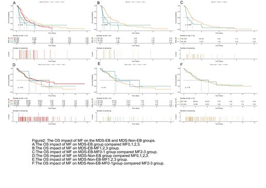
③The impact of MF on MDS-EB patients and MDS-Non-EB patients has different significances concerning OS, PFS and t-AML. As far as the OS prognostic factor, no matter the comparison of MF0, 1,2,3 four groups, the comparison MF1,2,3 three groups, the comparison of MF0-1 with MF2-3, all showed that MF was an adverse prognostic factor in the EB-MF group (All p value <0.05), and it also showed the adverse impact related with the grade of MF, but none of any in the MDS-Non-EB group (All p value>0.05)(Figure1). Comes to the PFS, it showed the MF on MDS-Non-EB was an inferior factor when compared MF+(MF1,2,3) to MF-(MF0)(p=0.0097), but not between the comparison MF1,2,3 groups(p=0.96) and none of the MDS-EB groups(p=0.31 and 0.092 respectively). The incidence of MF was related with a higher rate of t-AML not only in MDS-EB group but also in MDS-Non-EB group compared with MF0, no matter compared the MF+(MF1,2,3) with MF-(MF0) but also the comparison of MF0,1,2,3) (All P value < 0.001).
④ In the MDS-EB cohort, 66 patients in the MF-(MF-0) group completed Next-generation-sequencing (NGS) test, and 61 patients in the MF+ (MF1,2,3) group completed NGS testing. It seems that the incidence of gene mutation has no difference between the two groups. We conducted univariate COX regression analysis about the 30 genes (which were suggested in the NCCN guideline 2020) with OS, PFS and t-AML in the MDS-EB-MF cohort. The results showed that TP53, Nras, WT1 was related to the worse OS; U2AF1, Niras and TP53 was related to worse PFS and GATA2, IDH2, TP53, STAG2 was related to t-AML.
Conclusion:
MDS-MF has unique laboratory and clinical characteristics, act as an independent risk factor for shorter OS and worse PFS in the MDS-MF patients in our large cohort. In this study, we retrospectively analyzed 417 primary MDS cases in our hospital and analyzed the impact of MF on MDS-EB group and MDS-Non-EB group respectively. Our results showed the MF has a worse OS on the MDS-EB-MF group than the MDS-EB-MF0 group but no impact on MDS-Non-EB cohort. It was also consistent with the 5th edition of the WHO Classification of Haemato-lymphoid Tumors guidelines in which MDS-f was listed as a subtype concerning MDS with fibrosis concomitant with 5-19% blast cells in bone marrow or 2-19% blast cells in peripheral blood. The NGS sequencing showed TP53, Nras, WT1 was related to the worse OS; U2AF1, Niras and TP53 was related to worse PFS and GATA2, IDH2, TP53, STAG2 was related to t-AML.As far as we know, this is the first analysis of bone marrow fibrosis in MDS-EB patients. We need more research to help us understand the MDS-f subtype.
Disclosures
No relevant conflicts of interest to declare.


This feature is available to Subscribers Only
Sign In or Create an Account Close Modal